


A disease of non-infectious origin, in which the hyaline cartilage of the knee joint suffers first of all, which eventually collapses and ceases to perform its function, which further leads to the destruction of other components of the joint and leads to deformation.
This degenerative-dystrophic disease, as a rule, appears in women after 40 years, but men can also suffer, especially those who are overweight, prone to frequent hypothermia, involved in active sports or due to injuries.
Among all arthrosis, gonarthrosis of the knee joint is the most common.
There is an opinion that the cause of gonarthrosis is the deposition of salts in the joints. This opinion is absolutely wrong and the deposition of salts is rather a secondary process and causes pain during the development of the disease and is localized at the points of attachment of tendons and ligaments. Prevention plays an important role in disease prevention.
Anatomy of the knee joint
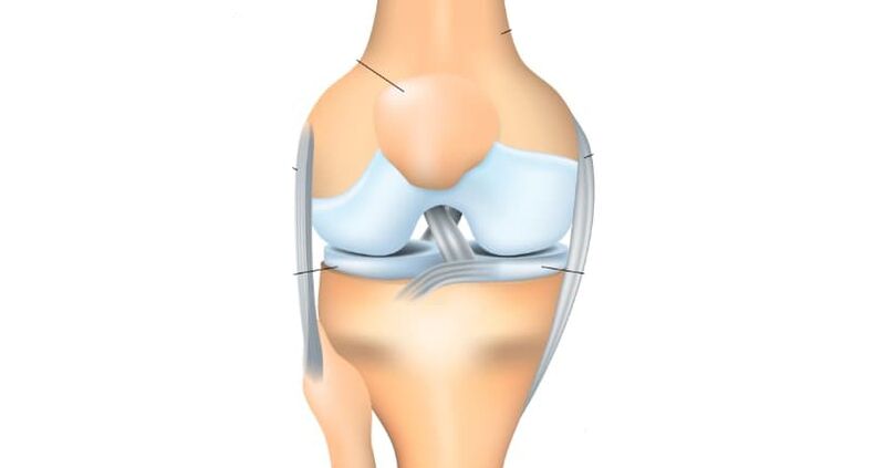
The knee joint consists of two surfaces, which are formed by the tibia and the femur. From the front, the knee joint protects the patella, which moves between the femoral condyles. The fibula does not participate in the formation of the knee joint and, in essence, does not carry any functional load, due to which it is often used to reconstruct other bony elements in the body.
All articular surfaces: tibia, femur and the inner surface of the patella are covered with hyaline cartilage, which is very smooth in structure, has a high degree of hardness and elasticity, the thickness of this dense and elastic structure reaches 5-6 mm. Cartilage absorbs shock during physical activity, prevents friction and softens impacts.
Classification of gonarthrosis
From the point of view of origin, gonarthrosis can be classified into primary, manifest, which occurs without injuries and secondary development, which is provoked by trauma, disease or developmental pathology and often appears as unilateral. In this case, the first type of gonarthrosis, as a rule, appears in the elderly and is rarely unilateral.
In its development, arthrosis of the knee joint goes through the following stages:
- The first stage of gonarthrosis- does not cause significant suffering to the patient, is characterized by constant pain or tightening pain, especially after heavy physical exercises, or direct load on the knee joint. The so-called "initial pain" symptom appears, when the patient stands up suddenly, painful sensations arise, which gradually disappear, but if an increased load is applied to the limb, the pain resumes. There may be slight swelling that goes away on its own. Rarely, but it happens, synovitis - fluid accumulates in the articular bag of the knee, due to which the knee area becomes spherical and swollen, movements in the limb are limited. At this stage, there is still no deformation of the joint.
- Second phase- the patient begins to be bothered by long and rather strong pains on the front and inner side of the joint, even with small loads, but after a long rest they usually disappear. When the joint moves, a crack is heard, if the patient tries to bend the limb as much as possible, a sharp pain appears. The amplitude in the movement of the joint is limited and the deformation begins to be detected. Synovitis appears often, bothers for a longer time, continues with a large accumulation of fluid in the joints.
- The third stage- causes considerable suffering to the patient, the pain is constant and bothers not only while walking, but also during rest and even at night, preventing sleep. The joint is already significantly deformed, the position of the limbs becomes X or O-shaped. A walking gait appears, and often, due to significant deformation, a person not only cannot bend, but also completely remove his leg, as a result of which he has to use a cane or even crutches to walk.
Pathology of gonarthrosis of the knee joint
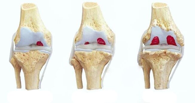
- In the initial, first stage of gonarthrosis, due to the development of a pathological process in the vessels that supply hyaline intraosseous cartilage, the articular surfaces gradually lose their inherent characteristics. They begin to dry, lose their smooth structure, cracks appear, due to which the gliding of the articular surfaces is disrupted, they begin to stick to each other, increasing surface defects. Hyaline cartilage degenerates, losing its shock-absorbing function due to continuous microtraumas.
- In the second stage of gonarthrosis, the degenerative-dystrophic manifestations increase: the joint space narrows, the articular surfaces flatten, adapting to the increasing loads. The part of the bone adjacent to the hyaline cartilage of the joint becomes denser and osteophytes appear along its edges, in the form of growth of bone tissue resembling spikes in shape. The capsule of the knee joint also undergoes changes, losing its elasticity. The fluid inside the joint becomes thicker and more viscous, changing its nutritional and lubricating properties, which further impairs joint function. Due to malnutrition, the condition of the hyaline cartilage worsens further, it begins to disintegrate and in some places it disappears completely. As a result of increased friction, the degeneration of the knee joint increases progressively, which leads to the third stage of gonarthrosis.
- In the third stage of gonarthrosis, a marked limitation of the range of motion in the joint appears. The surfaces are significantly deformed, hyaline cartilage is practically not present, the bones seem to be pressed into each other.
Reasons for the development of gonarthrosis
Basically, it is impossible to determine any single cause of gonarthrosis. Basically, its appearance is due to a combination of a number of reasons and a number of internal and external factors.
In 20-30% of cases, gonarthrosis is provoked by traumatic injuries to the knee joints or their components (ligaments, tendons, menisci), as well as fractures of the femur or tibia. The disease manifests, as a rule, 3-5 years after the injury. But there have been cases of the development of gonarthrosis in the early period (2-3 months).
In some patients, gonarthrosis can be caused by high physical exertion. Often, active physical activity can provoke a disease, especially after 40 years, when people begin to exercise actively to maintain health and understand the need for a healthy lifestyle. Above all, the load on the joints is when you run, as well as when you jump and sit.
Excess weight can also lead to the appearance of gonarthrosis, especially in combination with varicose veins of the lower extremities. The load on the knee joints increases and microtraumas or even serious injuries of the meniscus or the ligamentous apparatus of the joint occur. In this case, recovery is much more difficult, because. it is impossible to quickly lose excess weight in order to relieve the load on the joints.
Different types of arthritis (gout, psoriatic, rheumatoid, reactive or Bechterew's disease), some neurological pathologies (spinal injuries, craniocerebral injuries and other diseases that occur with damaged innervation of the lower extremities), as well as diseasesinherited, can provoke the development of gonarthrosis. causing weakening of the connective tissue.
Diagnosis of gonarthrosis
For the patient to be diagnosed with gonarthrosis, a combination of collecting complaints, examination and X-ray studies is necessary.
Today, an X-ray image of a joint is the simplest and most accessible research method, with the help of which it is possible to diagnose a patient with a sufficient degree of accuracy, observe the development of the process in dynamicsand determine further treatment tactics. Among other things, radiography allows you to make a differential diagnosis, for example, to exclude a tumor process in the bone tissue of the thigh or in the lower part of the leg or inflammatory. Also, for the diagnosis of gonarthrosis, computerized tomography and magnetic resonance are used, which can show changes not only in bone structures, but also in soft tissues.
In old age, everyone has certain signs of gonarthrosis, so the diagnosis can be made only after a complete collection of anamnestic data, complaints and visual examination, as well as instrumental research methods.
Treatment of gonarthrosis of the knee joint
When the first signs of a knee joint disease appear, it is necessary to consult an orthopedic doctor as soon as possible. In the initial stage of the process, the doctor prescribes drug therapy and complete rest of the affected limb.
After the end of the acute period, it is possible to appoint:
- exercise therapy course,
- massage,
- as well as physiotherapy procedures (electrophoresis with analgesics, UHF therapy, magnetic or laser therapy, phonophoresis with anti-inflammatory steroids, mud treatment, etc. )
In the next stage of treatment, the doctor can prescribe drug therapy, which includes taking chondroprotectors that stimulate metabolic processes in the joint. Sometimes intra-articular injections with drugs containing hormones are needed. If the patient has the opportunity to receive sanatorium-and-spa treatment, it is recommended for him. Often, to unload the joint, the patient is recommended to use a cane while walking. For prevention, you can use special orthopedic insoles or orthoses.
If the patient is diagnosed with the third stage of gonarthrosis, in which its manifestations are more pronounced (pain, damage or complete lack of joint function), surgical treatment may be required, which consists of knee arthroplasty. Rehabilitation measures until the function of the joint is fully restored, as a rule, last from 3 to 6 months, after which the patient can return to a normal life.
prevention
To avoid degenerative-destructive changes in the knee joint with age, it is necessary to resort to physical education, wear orthopedic shoes, control body weight and monitor the regime of rest and training.